United States Live Cell Imaging Market Report by Product (Equipment, Consumable, Software), Application (Cell Biology, Developmental Biology, Stem Cell and Drug Discovery, and Others), Technology (Time-Lapse Microscopy, Fluorescence Recovery after Photobleaching (FRAP), Fluorescence Resonance Energy Transfer (FRET), High Content Screening (HCS), and Others), and Region 2026-2034
Market Overview:
United States live cell imaging market size reached USD 676.2 Million in 2025. Looking forward, IMARC Group expects the market to reach USD 1,317.7 Million by 2034, exhibiting a growth rate (CAGR) of 7.46% during 2026-2034. The rising investments in R&D activities by pharmaceutical companies to assess drug efficacy and develop enhanced therapeutic solutions for patients are primarily propelling the United States live cell imaging market share.
|
Report Attribute
|
Key Statistics
|
|---|---|
|
Base Year
|
2025 |
|
Forecast Years
|
2026-2034
|
|
Historical Years
|
2020-2025
|
| Market Size in 2025 | USD 676.2 Million |
| Market Forecast in 2034 | USD 1,317.7 Million |
| Market Growth Rate (2026-2034) | 7.46% |
Access the full market insights report Request Sample
United States Live Cell Imaging Market Analysis:
- Major Drivers: The United States live cell imaging market growth is primarily driven by increasing pharmaceutical research and development (R&D) investments for drug efficacy assessment and therapeutic development. Advanced microscopy technologies, including super-resolution and light-sheet microscopy, enhance spatial resolution capabilities. Rising integration of artificial intelligence and machine learning accelerates data analysis and discovery processes.
- Key Market Trends: The expanding applications in drug discovery and development processes. Pharmaceutical companies increasingly rely on real-time assessment of drug efficacy and toxicity. Integration of AI-powered analysis systems enables efficient processing of vast imaging datasets, accelerating biomedical research outcomes.
- Market Challenges: High equipment costs and technical complexity limit widespread adoption among smaller research facilities. Maintaining optimal cellular conditions during prolonged imaging sessions presents ongoing technical challenges. Limited standardization across different imaging platforms creates compatibility issues for researchers and institutions seeking integrated solutions.
- Market Opportunities: Emerging applications in personalized medicine and precision therapeutics create significant growth opportunities. Expanding biotechnology sector and academic research investments drive United States live cell imaging market analysis toward promising prospects. Integration with advanced AI algorithms opens new possibilities for automated cellular behavior prediction and drug screening processes.
Live cell imaging stands as an important method in the scientific realm, enabling the observation of dynamic living cells within their growth medium as they undergo temporal transformations. The process involves numerous protocols and techniques, facilitating cell culture on the microscope stage while maintaining a consistent focal plane to track real-time cellular processes. Essential factors, such as imaging modality, media conditions, temperature, humidity, pH, osmolarity, photon dose, etc., influence the imaging outcomes, playing a crucial role in sustaining cellular health. Live cell imaging's versatility extends to monitoring cell fusion events, assessing molecular mobility through techniques like fluorescence recovery after photobleaching (FRAP), and gauging modifications such as growth or aging of condensate over time. It is indispensable for investigating fertilization, cellular development, signaling processes, etc., thereby providing invaluable insights to biologists, pharmacologists, and toxicologists.
United States Live Cell Imaging Market Trends:
Transformation through AI and ML Algorithms
The market is undergoing tremendous change through artificial intelligence integration and machine learning algorithms. These advances allow researchers to efficiently analyze large volumes of imaging data to detect cellular patterns and behaviors that cannot be detected manually. AI-based systems offer automatic cell tracking, morphology analysis, and predictive modeling functions. Machine learning algorithms enhance image quality, filtering noise out, and increasing data processing speeds. This technological innovation is especially useful in high-throughput screening contexts where thousands of cellular interactions need to be analyzed. Deep learning models incorporated into the technology allow decision-making in real-time during experiments, where the imaging parameters are optimized automatically. Sophisticated software solutions today provide cloud-based processing abilities so that researchers can remotely access powerful analytical tools. These developments lower analysis time considerably while enhancing accuracy and reproducibility of experimental outcomes between various research facilities.
Super-Resolution Microscopy and Enhanced Imaging Technologies
Breakthrough microscopy technologies are propelling United States live cell imaging market growth with record-breaking spatial and temporal resolution performance. Super-resolution methods, such as STED, PALM, and STORM microscopy, allow imaging of subcellular structures on nanometer scales unachievable with standard microscopy. Light-sheet fluorescence microscopy enables fast, gentle imaging of large biological samples with minimal photodamage. These sophisticated imaging modalities enable researchers to monitor dynamic cellular activities in real-time with unprecedented clarity and resolution. Multi-dimensional imaging capabilities record multiple fluorescence channels simultaneously, allowing detailed examination of intricate cellular interactions. Increased sensitivity detectors and advanced optical systems prolong imaging time while preserving cellular viability. The advancement of label-free imaging methods minimizes experimental artifacts and cellular disruption. Such technological innovations are especially useful in applications of developmental biology, neuroscience, and cancer research where precise analysis of cellular behavior is essential.
Expanding Drug Discovery and Pharmaceutical Applications
The increasing pharmaceutical applications and drug discovery procedures is aiding the market demand. Pharmaceutical industry is increasingly using live cell imaging for real-time drug efficacy, toxicity, and mechanism of action research. High-content screening platforms allow simultaneous examination of more than one cellular parameter, improving compound evaluation procedures. Live cell imaging gives essential information on drug kinetics, cellular uptake, and therapeutic response patterns. Personalized medicine uses patient-derived cellular models for drug selection and treatment optimization procedures. Oncology studies are especially enhanced by real-time observation of the behavior of tumor cells and monitoring of treatment response. Regenerative medicine applications involve live cell imaging to follow stem cell differentiation and tissue engineering. The technology facilitates the pharmaceutical industry to decrease development time and costs by early identification of potential therapeutic candidates and weeding out ineffectual compounds.
United States Live Cell Imaging Market Segmentation:
IMARC Group provides an analysis of the key trends in each segment of the market, along with forecasts at the country level for 2026-2034. Our report has categorized the market based on product, application, and technology.
Product Insights:
To get detailed segment analysis of this market Request Sample
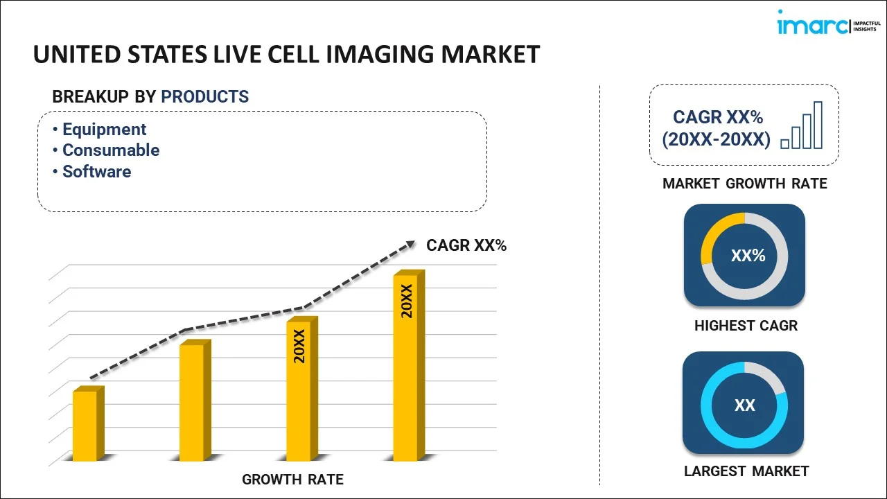
- Equipment
- Consumable
- Software
The report has provided a detailed breakup and analysis of the market based on the product. This includes equipment, consumable, and software.
Application Insights:
- Cell Biology
- Developmental Biology
- Stem Cell and Drug Discovery
- Others
A detailed breakup and analysis of the market based on the application have also been provided in the report. This includes cell biology, developmental biology, stem cell and drug discovery, and others.
Technology Insights:
- Time-Lapse Microscopy
- Fluorescence Recovery after Photobleaching (FRAP)
- Fluorescence Resonance Energy Transfer (FRET)
- High Content Screening (HCS)
- Others
The report has provided a detailed breakup and analysis of the market based on the technology. This includes time-lapse microscopy, fluorescence recovery after photobleaching (FRAP), fluorescence resonance energy transfer (FRET), high content screening (HCS), and others.
Regional Insights:
To get detailed regional analysis of this market Request Sample
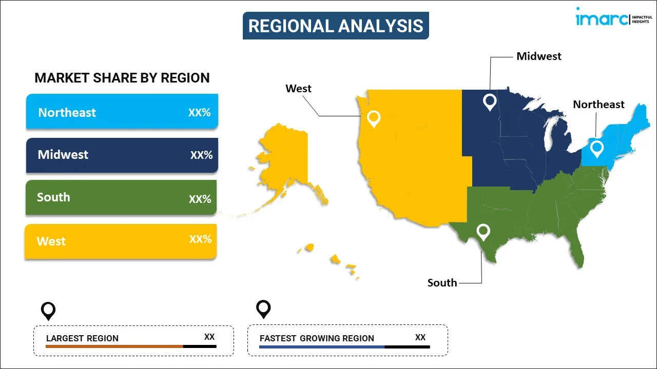
- Northeast
- Midwest
- South
- West
The report has also provided a comprehensive analysis of all the major regional markets, which include the Northeast, Midwest, South, and West.
Competitive Landscape:
The market research report has also provided a comprehensive analysis of the competitive landscape in the market. Competitive analysis such as market structure, key player positioning, top winning strategies, competitive dashboard, and company evaluation quadrant has been covered in the report. Also, detailed profiles of all major companies have been provided.
Recent News and Development:
- In January 2025, Durham-based Ramona launched Vireo™, the world’s fastest live-cell imaging system, designed to accelerate drug discovery. Using 24 miniaturized 4K microscopes and AI-powered real-time processing, Vireo analyzes a 96-well plate across five channels in under two minutes—far faster than traditional systems. It enhances throughput, reduces photodamage, and enables detailed cellular analysis. Early access at UNC and Carolina Institute validated its performance, and it will debut at SLAS 2025, showcasing advanced AI-driven imaging and analysis tools.
United States Live Cell Imaging Market Report Coverage:
| Report Features | Details |
|---|---|
| Base Year of the Analysis | 2025 |
| Historical Period | 2020-2025 |
| Forecast Period | 2026-2034 |
| Units | Million USD |
| Scope of the Report | Exploration of Historical and Forecast Trends, Industry Catalysts and Challenges, Segment-Wise Historical and Predictive Market Assessment:
|
| Products Covered | Equipment, Consumable, Software |
| Applications Covered | Cell Biology, Developmental Biology, Stem Cell and Drug Discovery, Others |
| Technologies Covered | Time-Lapse Microscopy, Fluorescence Recovery after Photobleaching (FRAP), Fluorescence Resonance Energy Transfer (FRET), High Content Screening (HCS), Others |
| Regions Covered | Northeast, Midwest, South, West |
| Customization Scope | 10% Free Customization |
| Post-Sale Analyst Support | 10-12 Weeks |
| Delivery Format | PDF and Excel through Email (We can also provide the editable version of the report in PPT/Word format on special request) |
Key Benefits for Stakeholders:
- IMARC’s industry report offers a comprehensive quantitative analysis of various market segments, historical and current market trends, market forecasts, and dynamics of the United States live cell imaging market from 2020-2034.
- The research report provides the latest information on the market drivers, challenges, and opportunities in the United States live cell imaging market.
- Porter's five forces analysis assist stakeholders in assessing the impact of new entrants, competitive rivalry, supplier power, buyer power, and the threat of substitution. It helps stakeholders to analyze the level of competition within the United States live cell imaging industry and its attractiveness.
- A competitive landscape allows stakeholders to understand their competitive environment and provides an insight into the current positions of key players in the market.
Frequently Asked Questions About the United States Live Cell Imaging Market Report
The live cell imaging market in the United States was valued at USD 676.2 Million in 2025.
The United States live cell imaging market is projected to exhibit a CAGR of 7.46% during 2026-2034, reaching a value of USD 1,317.7 Million by 2034.
The United States live cell imaging market is driven by advancements in microscopy technologies, rising demand for cell-based research, and increasing applications in drug discovery and cancer studies. Growing investment in life sciences, coupled with expanding academic and pharmaceutical research activities, further accelerates the adoption of live cell imaging solutions.
Need more help?
- Speak to our experienced analysts for insights on the current market scenarios.
- Include additional segments and countries to customize the report as per your requirement.
- Gain an unparalleled competitive advantage in your domain by understanding how to utilize the report and positively impacting your operations and revenue.
- For further assistance, please connect with our analysts.
 Request Customization
 Speak to an Analyst
 Request Brochure
 Inquire Before Buying
Benefits of Customization
- Personalize this research
- Triangulate with your data
- Get data as per your format and definition
- Gain a deeper dive into a specific application, geography, customer, or competitor
- Any level of personalization
Get in Touch With Us
UNITED STATES
Phone: +1-201-971-6302
INDIA
Phone: +91-120-433-0800
UNITED KINGDOM
Phone: +44-753-714-6104
Email: [email protected]
Client Testimonials


.webp)